Western Blot Imaging System: What Machine is Used for Western Blot?

Introduction
The Western Blot Imaging System has become an indispensable tool in various scientific fields, especially in molecular biology and clinical diagnostics. This system plays a critical role in detecting and analyzing specific proteins within a sample by utilizing sophisticated imaging technologies.
From research laboratories to biotech and clinical settings, Western blotting serves as one of the most powerful techniques for understanding protein expression, modifications, and interactions. With an increasing demand for accuracy and efficiency, selecting the right Western blot imaging system is crucial for obtaining reliable results.
In this article, we will explore the different components of the Western blotting process, what machines are used in Western blotting, and how to choose the best imaging solution for your specific needs.
This article will including below topics:
What is a Western Blot Imaging System?
What is Western Blot?
Western blotting is a widely used technique in molecular biology and biochemistry to detect specific proteins within a sample. The process involves several steps, including protein separation, transfer to a membrane, and detection.
Here’s a breakdown of the essential steps:
1. Protein Separation (Gel Electrophoresis):
Proteins are first separated according to their size using gel electrophoresis. This technique involves applying an electric field to a gel matrix, typically made of polyacrylamide, which causes proteins to migrate through the gel. Smaller proteins travel faster, while larger proteins move more slowly.
")
Image source: wiki.org
2. Protein Transfer:
After the proteins are separated, they are transferred from the gel onto a membrane (usually nitrocellulose or PVDF). This step is crucial for making the proteins accessible to antibodies that will be used for detection.

Image source: wiki.org
3. Protein Transfer:
The membrane is incubated with antibodies that specifically bind to the protein of interest. Once the antibody binds to its target protein, a detection method is used to visualize the protein. The most common detection methods are chemiluminescence, fluorescence, and near-infrared (NIR) imaging.

image source: wiki.org
The entire Western blotting process is instrumental in identifying proteins and studying their function, interaction, and modifications. However, the key to its success lies in the Western Blot Imaging System, which captures the final detection signals and allows for quantitative and qualitative analysis.
What is the Western Blot System?
A Western Blot System is the collection of equipment and reagents used to perform Western blotting. It includes:
- Electrophoresis Apparatus:
This is used for separating proteins by size using an electric field. It consists of a gel casting system, running buffer, and power supply. - Transfer System:
The transfer system moves the proteins from the gel to a membrane for further analysis. This system can use either wet or semi-dry transfer techniques. - Imaging System:
The imaging system is the crucial component that captures the signal emitted by the detection reagents (e.g., chemiluminescent substrates or fluorescent dyes). The captured image is then analyzed using specialized software.

image source: wiki.org
Together, these systems allow for detailed analysis of proteins, providing valuable insights into their expression, modifications, and interactions.
The Western Blot Imaging System, which uses a combination of light sources, detectors, and software, is responsible for converting the biochemical signals into readable data.
What Machine is Used for Western Blot?
Key Equipment for Western Blotting
1. Western Blot Imaging System:
This machine is the most critical piece of equipment when it comes to visualizing and quantifying proteins on the membrane. The imaging system captures the emitted light from the detection reagents and creates an image that can be analyzed. There are several different types of imaging systems available, each suited to different detection methods.
2. Electrophoresis System:
This system is responsible for separating proteins based on their size. The most used method is SDS-PAGE (sodium dodecyl sulfate-polyacrylamide gel electrophoresis). The system includes:
-
- Gel Casting Apparatus:
Used to prepare the gel matrix for electrophoresis. - Electrophoresis Chamber:
Where the gel is loaded with the protein samples and subjected to the electric field. - Power Supply:
Provides the necessary voltage for the electrophoresis process.
- Gel Casting Apparatus:
3.Transfer System:
After electrophoresis, the proteins must be transferred from the gel to a membrane. This can be done through:
-
- Wet Transfer:
Proteins are transferred from the gel to the membrane by applying an electric current in a buffer solution. - Semi-Dry Transfer:
A faster method where the gel and membrane are sandwiched between blotting papers and placed in a transfer buffer.
- Wet Transfer:
4. Detection Reagents:
These are chemicals that react with the proteins on the membrane to produce a detectable signal. The most common detection methods are:
-
- Chemiluminescence:
A light-emitting chemical reaction that generates light when the protein of interest reacts with a specific substrate. - Fluorescence:
Proteins are labeled with fluorescent dyes, and the emitted light is detected using specialized sensors. - Near-Infrared (NIR):
Proteins are labeled with NIR dyes, which can be detected using an NIR imaging system. NIR imaging offers the advantage of being less affected by background noise.
- Chemiluminescence:
Each of these components plays an essential role in the success of a Western blot experiment. The imaging system, however, is the final step in the process, converting chemical or light-based signals into digital images for analysis.


Applications for the Western Blot Imaging System
The Western Blot Imaging System is used in various fields, including:
1. Biomedical Research:
Western blotting is an essential tool for studying the expression and function of proteins in various biological systems. Researchers use Western blot imaging systems to explore cellular signaling, protein-protein interactions, and protein modifications (e.g., phosphorylation or acetylation).
2. Cancer Research:
Western blotting is often used in cancer research to identify biomarkers associated with cancer. The imaging system enables researchers to track the expression of cancer-related proteins and study the effects of potential treatments on these proteins.
3. Pharmaceutical Research and Drug Development:
Biotech and pharmaceutical companies use Western blotting to study drug targets and monitor the effects of drugs on protein expression. Western blot imaging systems help identify potential candidates for therapeutic development.
4. Clinical Diagnostics:
Western blotting is widely used in clinical diagnostics to detect and identify diseases associated with specific proteins. For example, it is used to confirm the diagnosis of HIV, Lyme disease, and various autoimmune diseases.
5. Immunology and Vaccine Development:
Western blot imaging systems are crucial in the study of immune responses. They are used to identify immune proteins such as antibodies and cytokines, making them vital in vaccine development and autoimmune disease research.

image source: wiki.org
How to Choose the Best Western Blot Imaging System?
Factors to Consider When Choosing a System
Selecting the right Western blot imaging system is essential to ensure accurate and reproducible results. Here are some critical factors to consider:
- Sensitivity:
High sensitivity is essential for detecting low-abundance proteins, particularly when working with complex samples like tissue lysates or serum. Choose a system with a high signal-to-noise ratio to detect faint signals. - Resolution:
The imaging system should have high resolution to distinguish between closely spaced protein bands. This is especially important when analyzing proteins of similar size. - Quantification Capabilities:
Many experiments require quantifying protein levels. Look for a system that allows for accurate quantification, ideally using software that supports standard curve generation and background subtraction. - Ease of Use:
A user-friendly interface is crucial for researchers who need to perform multiple experiments efficiently. Choose an imaging system with intuitive software that allows for easy analysis and data interpretation. - Cost and Budget:
The cost of the imaging system should align with the budget and the specific needs of the laboratory. While more advanced systems can be expensive, they often provide additional capabilities such as multiplexing or advanced quantification.
Chemiluminescence vs. Fluorescence vs. Near-Infrared: Which to Choose?
Chemiluminescence:
This is the most used detection method for Western blotting. It involves a chemical reaction that produces light, which is captured by the imaging system. It is highly sensitive but typically detects only one target protein at a time.

Immunoassay Methods and their Applications in Pharmaceutical Analysis:
Basic Methodology and Recent Advances. Int J Biomed Sci. 2006 Sep; 2(3): 217–235. PMID: 23674985.
image source:Darwish (2006).
Fluorescence:
Fluorescence-based detection is ideal for experiments where multiple proteins need to be detected simultaneously. Fluorescent dyes emit light when excited by specific wavelengths, allowing for the detection of multiple proteins in a single sample.

Figure : Indirect Fluorescent detection.
- A. Fluorescent dye-conjugated secondary antibody detects the primary antibody for the protein of interest.
- B. Light is used to excite the fluorescent conjugate at its excitation wavelength.
- C. The excited fluorescent dye emits light at its emission wavelength, which is detected via a digital imager, allowing visualization of the protein of interest.
Near-Infrared:
NIR detection is highly sensitive and offers a wide dynamic range, making it suitable for quantitative applications. NIR imaging is less affected by background signals and can be used for multiplexing with less interference.

Western blot using an anti-lipoic acid primary antibody and an IR-dye labelled secondary antibody in Leishmania major extracts.
(Source:wikimedia.org)
Recommendations Based on Specific Needs
- Research Laboratories:
For basic research, a chemiluminescent system is often sufficient. If multiplexing is needed, a fluorescence-based system would be more suitable. - Biotech and Pharmaceutical Companies:
For drug discovery and high-throughput screening, near-infrared or fluorescence-based imaging systems offer the ability to perform multiple assays simultaneously and with higher sensitivity. - Clinical Diagnostics:
Clinical labs require precision and reliability. A system with near-infrared detection is ideal for quantification and reproducibility.
Compare Western Blot Imaging Systems
Western blot imaging systems vary in capabilities, from fundamental chemiluminescent systems to advanced multimodal and near-infrared (NIR) imaging platforms. While newer technologies offer additional features, the standard chemiluminescent imaging system remains a cornerstone of protein detection due to its reliability, cost-effectiveness, and widespread adoption in research laboratories.
- Standard Chemiluminescent Imaging System:
Basic chemiluminescent system, suitable for standard single-protein detection. Simple, reliable, and cost-effective. - Multimodal Imaging System:
Hybrid system with both chemiluminescence and fluorescence capabilities. Ideal for labs that need to detect multiple proteins in one experiment. - Near-Infrared (NIR) Imaging System:
Near-infrared imaging system, providing high sensitivity and quantification capabilities. Best suited for clinical and pharmaceutical applications.
Comparison Table of Western Blot Imaging Systems
| System Type | Standard Chemiluminescent Imaging System | Multimodal Imaging System | Near-Infrared (NIR) Imaging System |
|---|---|---|---|
| Detection Methods | Chemiluminescence | Chemiluminescence + Fluorescence | Near-Infrared (NIR) Fluorescence |
| Best For | Foundational protein detection, academic and clinical research | Labs requiring multi-protein detection | Clinical, pharmaceutical, high-sensitivity research |
| Key Advantages | Cost-effective, highly sensitive, widely validated, and established in regulatory environments | Supports multiple detection methods, increased flexibility | High sensitivity, strong quantification, minimal background noise |
| Considerations | Requires chemiluminescent substrate, limited multiplexing | Higher cost, may require specialized reagents | Higher initial investment requires NIR-compatible dyes |

Why the Standard Chemiluminescent Imaging System Remains Essential
(Source:wikimedia.org)
The Standard Chemiluminescent Imaging System continues to be a critical tool in life science research. It is the most widely used and validated method for protein detection, making it the gold standard for labs around the world.
While newer imaging technologies provide additional functionalities, chemiluminescence remains the foundation of Western blot imaging, offering:
- Exceptional Sensitivity: Detects low-abundance proteins with high specificity.
- Cost-Effectiveness: More affordable compared to fluorescence or NIR-based systems, making it accessible for academic labs and smaller research institutions.
- Broad Compatibility: Well-established protocols, reagents, and workflows make it easy to integrate into existing research setups.
Regulatory and Industry Acceptance: Frequently used in regulated environments, ensuring data reproducibility and compliance with scientific standards.
Choosing the Right System
For laboratories conducting routine Western blotting, a Standard Chemiluminescent Imaging System is often the best choice due to its reliability and accessibility. However, for research that requires multiplexing or advanced quantification, a Multimodal or Near-Infrared Imaging System may provide additional benefits.
The choice depends on research objectives, budget, and the level of complexity required for protein analysis.
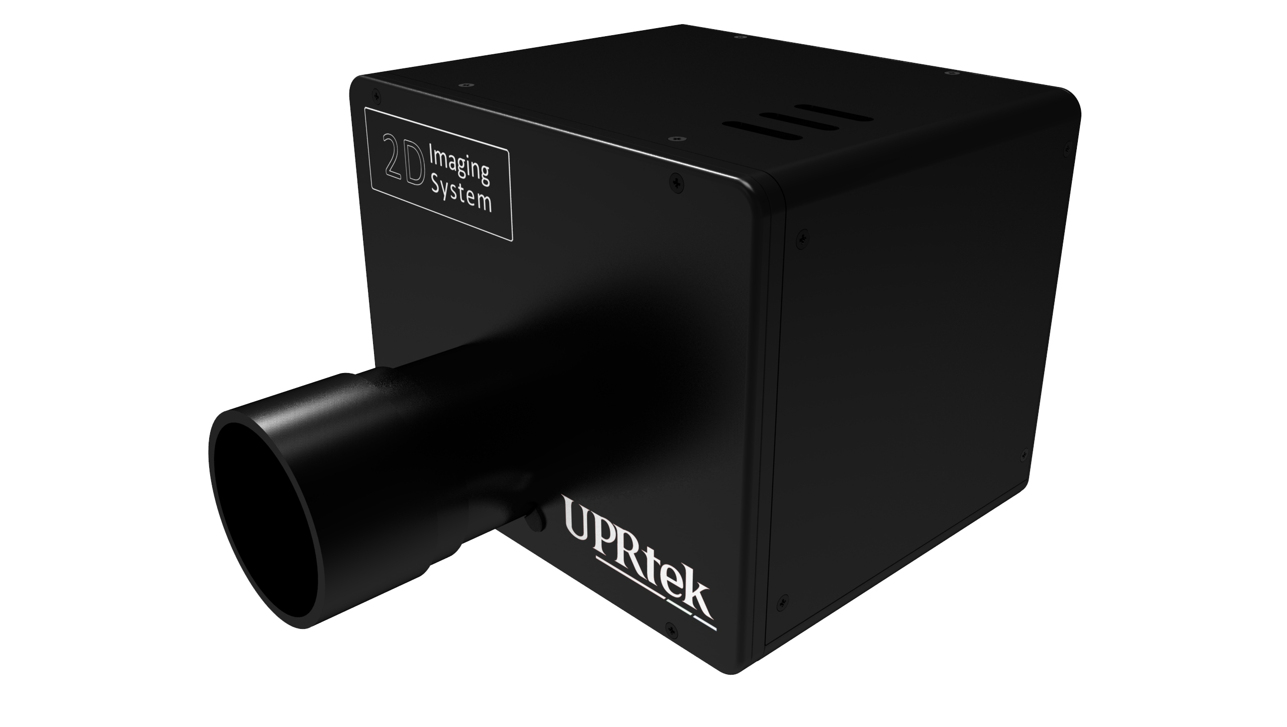
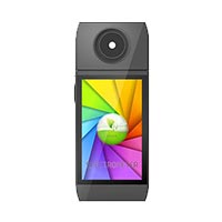
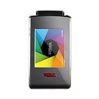
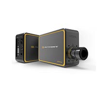
Western Blot Imaging Solutions – Why Choose UPRtek?
UPRtek’s Innovative Approach to Western Blot Imaging
Many laboratories face challenges such as high costs, complex data analysis, and the need for high sensitivity when performing Western blotting.
The right imaging system can address these issues by delivering consistent, accurate, and efficient results tailored to the needs of researchers.
UPRtek’s Technological Expertise & Customization Capabilities
UPRtek specializes in high-performance Western blot imaging solutions, offering a patented, cost-effective, and flexible chemiluminescent system that operates as a standalone unit.Designed for ease of use and accessibility, it provides research, biotechnology, and clinical laboratories with a streamlined and reliable imaging solution for fundamental Western blotting needs.
While UPRtek’s current offerings focus on chemiluminescence, our proven track record in global project development enables us to rapidly innovate and customize advanced systems—including fluorescence and near-infrared imaging—based on customer requirements.
With extensive experience in optical system engineering and precision measurement, UPRtek is fully capable of delivering tailored solutions that evolve with scientific advancements and laboratory demands.
By combining technical expertise, innovative engineering, and a commitment to customization, UPRtek ensures that laboratories have the right tools to achieve high-quality imaging and data accuracy, while maintaining flexibility for future advancements.
Conclusion
The Western Blot Imaging System plays a fundamental role in protein detection and analysis, supporting critical research in fields such as molecular biology, biotechnology, and clinical diagnostics.
However, despite its widespread use, the core technology behind Western blot imaging has seen relatively slow advancements over the years. Traditional imaging systems often focus solely on incremental improvements in sensitivity and resolution, but they have lacked breakthrough innovations in automation, integration, and usability.
The evolution of optical imaging in this domain has been gradual, with limited progress in enhancing workflow efficiency, improving accessibility, and reducing costs.
This is precisely where UPRtek excels. By leveraging its expertise in optical engineering, precision measurement, and system integration, UPRtek has redefined the Western Blot Imaging System with a more flexible, cost-effective, and standalone solution that meets the fundamental needs of researchers while laying the groundwork for future enhancements.
With patented optical technology and a commitment to innovation, UPRtek is uniquely positioned to bridge the gap between traditional Western blot imaging and modern scientific demands.
Furthermore, as the industry gradually moves toward smarter, more efficient laboratory workflows, technologies such as AI-assisted analysis, automated exposure control, and enhanced imaging software are becoming increasingly essential.
While many existing systems struggle to integrate these advancements, UPRtek has the technical expertise and global development experience to push these boundaries. Although its current product line focuses on chemiluminescence-based imaging, UPRtek has the capability to rapidly develop and customize advanced solutions, including fluorescence and near-infrared imaging, based on market demand.
In a field where innovation has been slow, UPRtek is leading the charge by introducing new possibilities in Western blot imaging—bringing a fresh perspective to a technology that has remained largely unchanged for years.
By offering flexibility, precision, and the ability to evolve with scientific advancements, UPRtek is setting a new standard for Western blot imaging, ensuring that researchers and laboratories have the right tools to achieve highly accurate and reliable results, both now and in the future.
Hot Product
Handbook Series

The Flicker Handbook
Everything thing you need to know about Flicker, an insidious, potentially serious lighting artifact impacting visual safety for public places like hospitals, offices, libraries, and more...
About UPRtek
United Power Research and Technology
UPRtek (est. 2010) is a manufacturer of portable, high-precision light measurement instruments; Handheld Spectrometers, PAR meters, Spectroradiometers, Light Calibration Solutions.
UPRtek HQ, R&D and manufacturing are all based out of Taiwan, with Worldwide representation through our certified Global Resellers.

UPRTEK AT TOUCH TAIWAN 2025 – DISCOVER THE LATEST IN LIGHT MEASUREMENT TECHNOLOGY
Join Us at Booth L412 | April 16-18, 2025 | Taipei Nangang Exhibition Center
Latest Articles
Category

0 Comments